Figure 1

Figure 2

Biomedical Engineering
Team 27
Team Members |
Faculty Advisor |
Tudor Ilies |
Bin Feng Sponsor Medtronic |
sponsored by

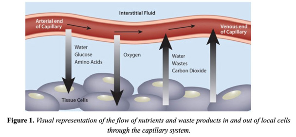
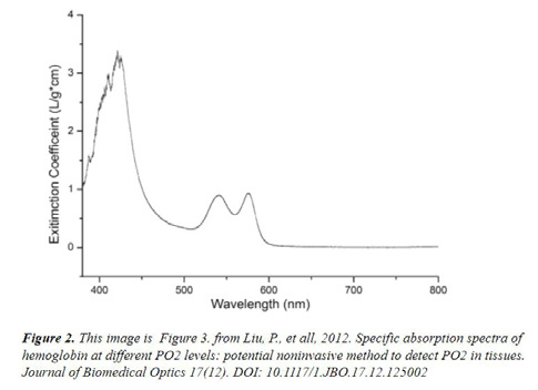
Minimally invasive surgery is a highly advantageous, up and coming field of surgery that limits the number and size of incisions made on the patient, and has proven to be much safer than open surgery. It has been found that adequate local tissue perfusion at the tissue incision site contributes to the success rate of surgical procedures. Being able to identify and quantify local tissue perfusion prior to making an incision would help surgeons operate on the healthiest tissue that is most likely to successfully heal. The goals of this project were to explore various methods of tissue perfusion detection and complete a proof of concept study demonstrating the application of UV-VIS fiber optic spectrometry utilizing two wavelength ranges ( ultra-violet and visible) for local tissue perfusion. A visual representation of local perfusion is shown in Figure 1. The graph shown in Figure 2. is a graph of the unique absorbance peaks of hemoglobin over the utra-violet to visible light wavelength range.
