Figure 1
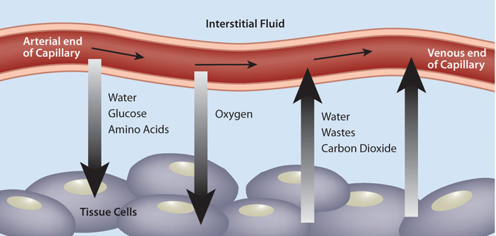
Figure 2
Team 21
Team Members |
Faculty Advisor |
Emily Shea |
Bin Feng Sponsor Medtronic |
sponsored by
![]()
Minimally invasive surgery is a growing branch of the medical field, concerned with operations that lead to decreased trauma to the body. Laparoscopy for example, uses one or more small incision to allow surgical instruments to access the body with the least amount of damage to the tissue. Minimally invasive surgery is a growing field due to its many advantages in comparison with open surgery. It decreases the patient's risk for infection, increases healing rates, physically there is less bleeding and less scarring, and this is all followed by less hospital time. An important component of surgical procedures is the perfusion of biological tissue. Tissue perfusion can be defined as local blood flow through a capillary bed within a tissue. It has been found that good local tissue perfusion helps the success rate and healing of a surgical procedure. This project aims to find a way to quantify local perfusion of tissue. In doing so, the goal is that tissues can be identified as well-perfused or ill-perfused, which would help a surgeon determine the ideal location for incisions. Perfusion can be quantified by the oxygenation of the blood (more specifically the hemoglobin) in a tissue. The overall goal of this project is to evaluate a state-of-the-art perfusion detection method and complete a proof-of-concept study demonstrating the application of UV-Vis Fiber Optic Spectrometry for local tissue perfusion.
